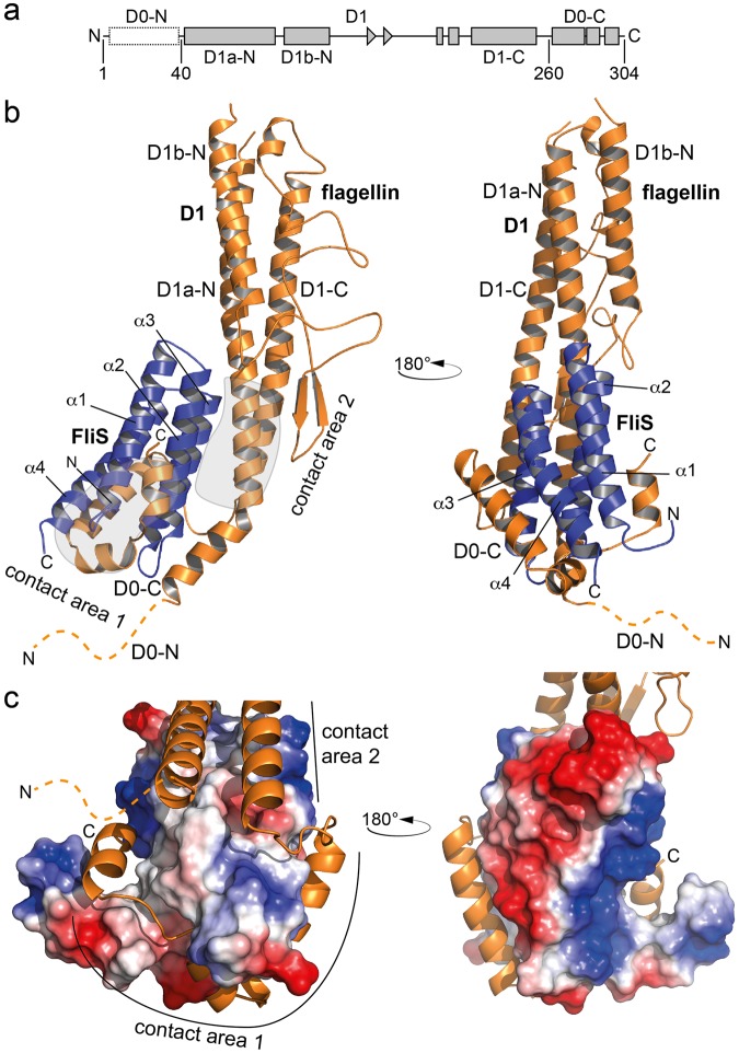
Figure 1.
Crystal structure of the flagellin/FliS heterodimer. (a) Domain organization of flagellin. (b) Crystal structure of flagellin/FliS as a cartoon representation in two orientations. flagellin and FliS are shown in orange and blue, respectively. ‘N’ and ‘C’ indicate N- and C-termini, respectively. The disordered N-terminus of flagellin is shown as dashed line. Contact areas between flagellin and FliS are encircled in grey. (c) Electrostatic surface potential of FliS within the contact area of flagellin to FliS. The flagellin-binding pocket is of hydrophobic and polar nature (left side), whereas the opposite site of FliS is highly charged (right side).

