Figure 3.
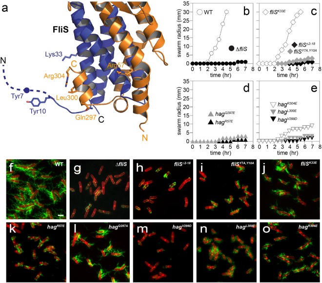
Physiology of flagellin/FliS binding to FlhA-C. (a) Projection of different amino acid exchanges onto the structure of flagellin/FliS. Flagellin is shown in orange, while FliS is depicted in blue. Dashed lines indicate disordered regions. (b–e) Quantitative swarm expansion assay for fliS and flagellin (hag) mutants: WT is swarming proficient and covers a 0.7% LB agar plate in about 4.5 hours but ΔfliS is non-swarming. Both fliSΔ2–18 and fliSY7A,Y10A phenocopy a ΔfliS mutant for swarming. fliSK33E is swarming proficient. hagR57E confers a non-swarming phenotype while all hag mutants except hagV299D show almost no swarming motility. (f–o) Fluorescence microscopy of B. subtilis filaments shows deficiency of ΔfliS, fliSΔ2-18 and fliSY7A,Y10A mutants to secrete flagellin. fliSK33E shows short flagellar filaments. hagR57E, hagV299D, hagL300E and hagR304E show almost no flagellar filaments, while hagQ297A exhibits short flagellar filaments. Scale bars are 2 µm.

