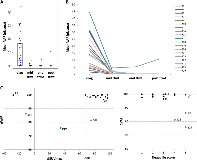
Fig. 5. Longitudinal assessment of mutation abundance in plasma cfDNA upon R-CHOP treatment according to interim PET scan.
a Distribution of the cfDNA mean VAFs of the patients at the different times of follow-up (diagnosis, mid-treatment, end of treatment, and 6 months post-treatment). b Evolution of the cfDNA mean VAFs for each patient throughout treatment. c ΔVAF values in plasma according to the ΔSUVmax (left) or Deauville score (right) between diagnosis and mid-treatment. The vertical dashed lines represent the cut-off ΔSUVmax of 70% (left) or Deauville score of 3 (right), and the blue dots represent patients with ΔVAF <90%

