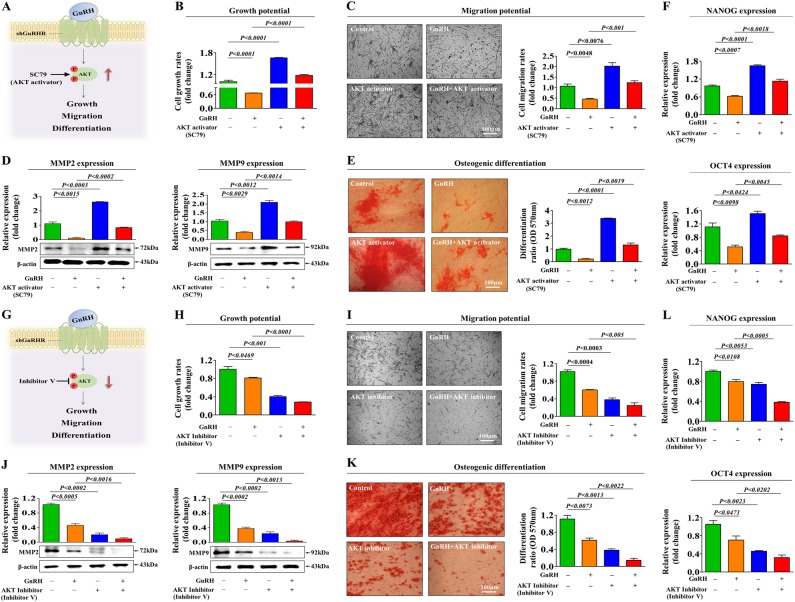
Fig. 4. Activation or inhibition of Akt signaling attenuates the GnRH-induced effects on multiple stem cell functions.
Schematic representation of the experimental protocol as described in the materials and methods section (a). Endometrial stem cells were pretreated with the Akt activator SC79 (10 µM) for 24 h prior to treatment with 1 µM GnRH for 48 h, and the changes in cell viability were determined by an MTT assay. Stem cell viability (%) was calculated as a percent of the vehicle control (b). The changes in migratory capacity were measured via the transwell assay (c) and western blotting for MMP-2 and MMP-9 (d). The changes in osteoblast differentiation were determined by alizarin red staining. The relative quantification of calcium mineral content was performed by measuring the absorbance at 570 nm (e). The ability of SC79 to attenuate the GnRH-induced inhibition of stem cell marker expression (NANOG and OCT4) was determined by real-time PCR (f). Schematic representation of the experimental protocol as described in the materials and methods section (g). Endometrial stem cells were pretreated with Akt inhibitor V (10 µM) for 24 h prior to treatment with 1 µM GnRH for 48 h, and the changes in cell viability were determined by an MTT assay. Stem cell viability (%) was calculated as a percent of the vehicle control (h). The changes in migratory capacity were measured via the transwell assay (i) and western blotting for MMP-2 and MMP-9 (j). The changes in osteoblast differentiation were determined by alizarin red staining. The relative quantification of calcium mineral content was performed by measuring the absorbance at 570 nm (k). The synergism between Akt inhibitor V and GnRH in inhibiting stem cell marker expression (NANOG and OCT4) was determined by real-time PCR (l). β-actin was used as the internal control. The data are presented as the mean ± SD of three independent experiments

