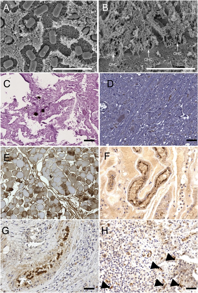
Figure 2.
Microscopic findings. Some of the most relevant microscopic findings revealed by scansion electron microscopy (SEM), histopathology, and immunohistochemistry (IHC) examinations: (A) evidences of post-mortem bacteria proliferation and (B) post-mortem degeneration of Organo del Corti outer hear cells (OHC, white arrows) observed at SEM. (C) occasional fat emboli within pulmonary capillary vessels of SW3 revealed by OsO4 post-fixation technique, PAS 20×. (D) Positive immunostaining of axons using anti-caspases-3 antibody, suggesting ongoing apoptotic changes in in SW2’s brain; 40×. (E) IHC on muscular tissues revealed a multifocal cytoplasmic immunostaining within fibers by using anti-fibrinogen antibody suggesting an ongoing damage (40×); (F) rabdomyolisis was further confirm by IHC on kidneys using anti-myoglobin antibody: the picture shows myoglobin granules in the apical side of tubular epithelial cells (40×); (G) positive IHC staining of circulating monocytes in the spleen of SW3 by using anti-CDV antibody (40×); (H) this image shows the same results of the previous image in dendritic cells of SW3’s spleen (arrows), 40×.

