Figure 2.
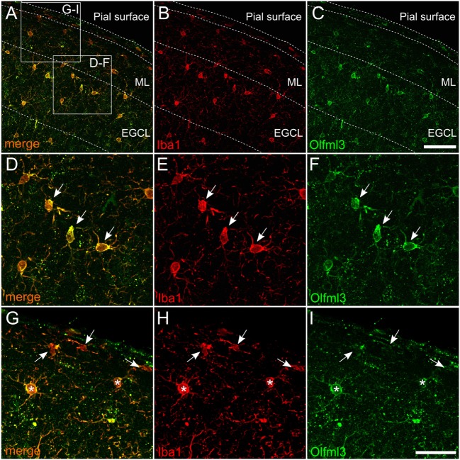
Expression of Olfml3 in cortical microglia. Overlay (A) of Iba1+ (B) and Olfml3+ (C) cortical microglia from 6-month-old C57BL/6 mice demonstrate microglia-specific expression of Olfml3. White arrows in high magnification images show strong cytoplasmic immunoreactivity for Olfml3 (F) in Iba1+ microglia (D,E). Noteworthy, Iba1+ meningeal macrophages (white arrows) located in close proximity to the pial surface show faint Olfml3 expression as compared to microglia (white asterisks) of the ML (G–I). Abbreviations: ML, molecular layer; EGCL, external granule cell layer. Scale bars represent 20 µm (A–C) and 10 µm (D–I).

