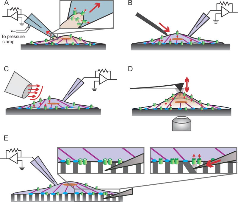
Fig. 2.
Piezo1 transduces outside-in mechanical forces. A variety of techniques have been developed to study transduction of outside-in mechanical forces by Piezo1. Some of these include: A) Membrane stretch elicited by suction pulses imparted by a high-speed pressure clamp in cell-attached patch clamp mode. B) Membrane stretch elicited by cell indentation with a glass probe controlled by a piezoelectric actuator in whole-cell patch clamp configuration. C) Shear stress induced by pulses of fluid flow from a perfusion pipette in whole-cell patch clamp configuration. D) Pulling or pushing on the cell surface by an AFM cantilever, while channel activity is measured by Ca2+ imaging on a confocal microscope. E) Cells are seeded on an array of microposts; a glass probe mounted on a piezoelectric actuator deflects a single micropost, mechanically stimulating a small number of channels in the vicinity of the micropost, while electrical activity is measured with whole-cell patch clamp. See Section 3 of the text for details on the techniques and results obtained. In all panels, actin filaments are shown in purple, focal adhesion zones in blue, Piezo1 molecules in green. Solid red arrows indicate force application; small broken arrows indicate ionic conduction through the channel.

