Fig. 3.
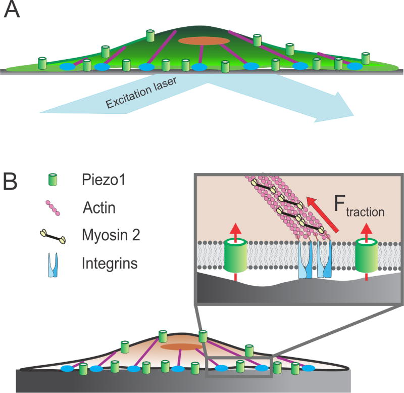
Piezo1 transduces inside-out mechanical forces. A) Activation of Piezo1 by inside-out forces is studied by imaging Ca2+ influx through the channel with TIRFM. Since forces are generated by the cell itself, no external force stimulus is applied to the cell. B) Piezo1 is activated by traction forces (solid red arrow) generated at integrin-rich focal adhesion zones (blue) by myosin 2 molecules (black and yellow) along the actin cytoskeleton (purple). Broken red arrows denote ionic conduction through the channel. See Section 3 for details.

