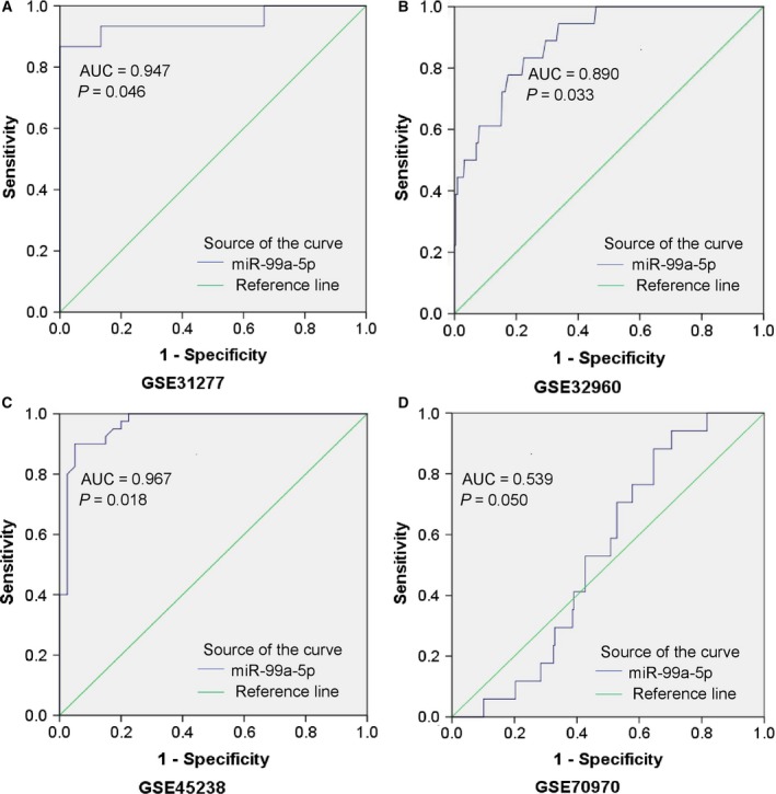
Figure 8.

Representative ROC curves of the microarrays. ROC curve of miR‐99a‐5p expression in normal and HNSCC tissues from microarrays with P value ≤ 0.05 were plotted: (A) http://www.ncbi.nlm.nih.gov/geo/query/acc.cgi?acc=GSE31277 (AUC = 0.947, P = 0.046). (B) http://www.ncbi.nlm.nih.gov/geo/query/acc.cgi?acc=GSE32960 (AUC = 0.890, P = 0.033). (C) http://www.ncbi.nlm.nih.gov/geo/query/acc.cgi?acc=GSE45238 (AUC = 0.967, P = 0.018). (D) http://www.ncbi.nlm.nih.gov/geo/query/acc.cgi?acc=GSE70970 (AUC = 0.539, P = 0.050).
