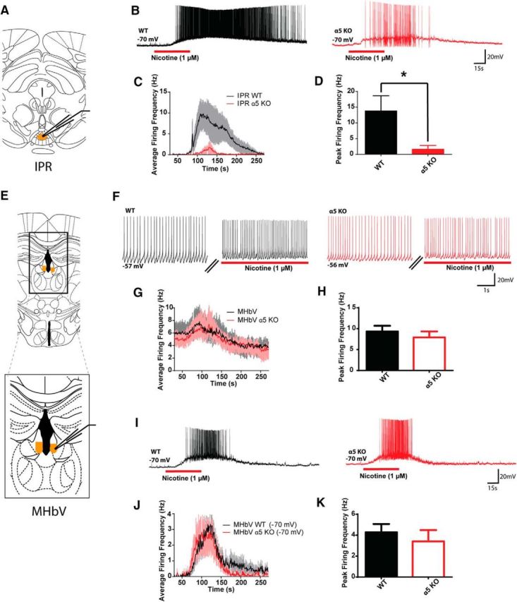
Figure 2.

Assessing the effects of nicotine on neuronal excitability in the MHbV and IP in WT and α5KO mice. A, Schematic of anterior recording site for IP neurons. B, Example traces of IPR neuron responses to nicotine in WT and α5KO mice. C, Average IPR neuronal firing frequency across baseline, nicotine (1 μm, 1 min), and washout recording. D, Peak nicotine-elicited firing frequency is significantly larger for WT than α5KO IPR neurons (Mann–Whitney U = 46.0, p = 0.02). E, Schematic illustrating recording sites for MHbV neurons. F, Example traces of MHbV neuron responses to nicotine at resting membrane potential in WT and α5KO mice. G, Average MHbV neuronal firing frequency across baseline, nicotine (1 μm, 1 min) and washout recording. H, Peak nicotine-elicited firing frequency does not differ between WT and α5KO mice in MHbV neurons at resting membrane potential (Mann–Whitney U = 49.0, p = 0.2). I, Example traces of MHbV neuron responses to nicotine in WT and α5KO mice recorded from −70 mV. J, Average MHbV neuronal firing frequency across baseline, nicotine (1 μm, 1 min) and washout recording from −70 mV. K, Peak MHbV neuronal firing frequency does not differ between WT and α5KO mice recorded from −70 mV (Mann–Whitney U = 58.0, p = 0.3). See Figure 2-1 (in table form). *p < 0.05.
