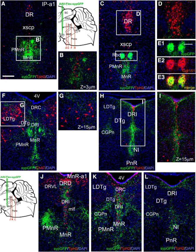
Figure 7.

Efferents of Chrna5-expressing neurons to the pontine raphe and tegmentum. A–I, Caudal projections of IPR neurons labeled with Cre-dependent AAVs encoding a red marker in cell bodies and axons (tdTomato) and a green marker targeted to presynaptic areas (sypGFP), as shown in Figure 5A. A, B, Synaptic labeling in the MnR/PMnR (bregma 4.2). Serotonergic neurons are marked by immunofluorescent staining for Tph2. Confocal image in B shows that labeling surrounds but usually does not overlap Tph2-expressing cell bodies. C, D, Synaptic labeling in the rhabdoid nucleus and MnR/PMnR (bregma 4.5). Confocal view shows that fibers are very sparse in the DR. 5HT neurons are identified by immunostaining for Tph2. E, Coimmunostaining for Gpr151 and sypGFP in the rhabdoid nucleus. F, G, Synaptic labeling in the caudal MnR/PMnR, DR, and dorsal tegmentum (bregma 5.0). Cholinergic neurons are marked by immunofluorescent staining for ChAT. Sparse sypGFP labeling is noted in the LDTg, which does not overlap the ChAT-expressing neurons. H, I, Synaptic labeling in the pontine tegmentum (bregma 5.3). Synaptic labeling is prominent in DRI and NI, but absent from DTg and PnR. J–L, Caudal projections of Chrna5Cre-expressing MnR neurons in case MnR-a1, labeled as shown in Figure 5H. J, Prominent synaptic labeling in DRI and in PMnR ipsilateral to the injection (bregma 4.8). K, Labeling in CGPn and DRI sparing DTg/LDTg (bregma 5.0, comparable to F). L, Projections to the caudal part of the pontine tegmentum, including NI (bregma 5.3, comparable to H). MnR, Median raphe nucleus; PMnR, paramedian raphe nucleus; Rbd, rhabdoid nucleus; mlf, medial longitudinal fasciculus; xscp, decussation of the superior cerebellar peduncle; DTg, dorsal tegmental nucleus; LDTg, laterodorsal tegmental nucleus; PnR, pontine raphe nucleus; NI, nucleus incertus; CGPn, central gray of the pons; DR, dorsal raphe nucleus; DRI, dorsal raphe nucleus, interfascicular part; DRD, dorsal raphe nucleus, dorsal part; DRVL, dorsal raphe nucleus, ventrolateral part; DRC, dorsal raphe nucleus, caudal part. Scale bar, 200 μm.
