
Figure 9.
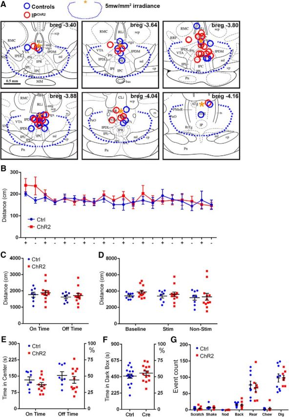
Activation of Chrna5Cre neurons does not affect locomotion, produce acute anxiety, or elicit somatic signs of nicotine withdrawal. A, Placement of fiber-optic cannulas in IPChR2 and control mice. Data for three behavioral cohorts of mice are shown. Blue circles represent fiber placement in control mice; red circles represent placement in IP ChR2 mice. Fiber termini are shown on images from a standard atlas closest to their rostrocaudal position (Paxinos and Franklin, 2001). In all cases, the optical fibers were intact and transmitted light efficiently when examined postmortem after the experimental protocol. A small number of cannulas were located outside the predicted area for effective illumination of IPR, such as the most dorsal cannula at bregma −3.8 and the most dorsal two cannulas at bregma −4.16, but the intensity of target illumination cannot be precisely determined. To avoid selection bias, all subjects were included in the statistical analysis except for two subjects in which the cannula placement obstructed the aqueduct and induced hydrocephalus (data not shown). The blue dashed line indicates the limit of 5mw/mm2 irradiance, as measured from the point indicated by the yellow asterisk. B–E, Open-field behavior was assessed in IPChR2 and control mice over three 10 min intervals: baseline, stimulation, and poststimulation. The stimulation period consisted of 30 s periods of 20 Hz light pulses, alternating with 30 s periods of lights off. B, Distance traveled during 30 s intervals of pulsed light (+) and no light (−) during the stimulation period. Cohort of 12 IPChR2 and 8 control mice. C, Summary of distance traveled during light on time and light off time during the stimulation period; no effect of light or genotype was observed. D, Summary of distance traveled during the baseline, stimulation, and poststimulation periods. No effect of period or genotype was observed. E, Time spent in the center of the open-field enclosure during the stimulation period. No effect of light or genotype was observed. F, Time spent in the dark chamber of the light/dark box; no effect of light or genotype was observed. Cohort of 14 IPChR2 and 16 control mice. G, Somatic signs of nicotine withdrawal. Intermittent pulsed stimulation was delivered to the IP of nicotine-naive IPChR2 and control mice (see Materials and Methods) and mice were assessed for six somatic signs of nicotine withdrawal. No effect of genotype was observed. Cohort of 8 IPChR2 and 9 control mice. Cli, Caudal linear nucleus raphe; MnR, median raphe; PMnR, paramedian raphe; Pn, pons; PnO, pontine reticular nucleus, oral; PN, paranigral nucleus; RLi, rostral linear nucleus raphe; RRF, retrorubral field; SNc, substantia nigra, pars compacta; SNr, substantia nigra, pars reticulata. Scale bar, 0.5 mm. For extended data regarding cannula placement, see Figure 9-1. Pulsed light stimulation induced the expression of cFos in the IP in a pattern similar to that previously obtained with systemic nicotine activation (Ren and Sagar, 1992). For extended data on cFos induction by optogenetic stimulation, see Figure 9-2.