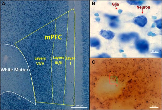
Figure 2.
A, Parcellation of layers I, II/III, and V/VI of the mPFC, as well as the underlying white matter, in a Methylene Blue/Azure II section. B, Higher magnification of a Methylene Blue/Azure II section in which neurons can easily be distinguished from glia. C, High magnification of an immunohistochemically stained section of synaptophysin, a marker of synapses. The counting frame has green inclusion and red exclusion lines for stereological counting.

