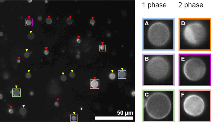
Figure 4: assigning phase state of individual vesicles within a population imaged at fixed temperature.

(Left) An example image showing a field of GPMVs in which most vesicles can be unambiguously assigned to contain either 1 or liquid 2 phases. Yellow triangles point towards 1 phase vesicles while red triangles point towards 2 phase vesicles. (right) isolated view of selected vesicles shown at the Left. GPMVs shown in A-C contain a single phase. A and B show GPMVs where the image is focused on a vesicle surface, and the brightness of the fluorophore is uniform across this surface. The GPMV in C is in focus along the midplane of the vesicle, and the brightness of fluorophore is uniform around the circumference. GPMVs shown in D and E contain coexisting liquid phases. The vesicle in D where the image is focused on the vesicle surface and a clear phase boundary is observed. The vesicle in E is focused close to the midplane, and the fluorophore intensity varies along the perimeter. F is difficult to assign. However, both the surface and the circumference vary in brightness so it was classified as containing two phases. In this example, some GPMVs in the field were not labeled because they are too small or too out of focus. In general, it is most important to remain consistent regarding assignments and the characteristics of vesicles that the investigator chooses not to assign. One reason to image at least 100 vesicles is to reduce the possible contributions of user bias in assigning vesicles.
