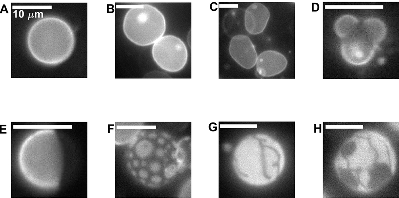
Figure 5: Images of vesicles highlighting the range of possible shapes and domain morphologies.

(A) Spherical vesicle with a single liquid phase. (B) Two single phase vesicles that each contain internal smaller vesicles (bright spots) . (C) Non-spherical vesicles imaged above Tmix. Vesicle edges undulate when vesicles are imaged in time (not shown). (D) Vesicle with bulging liquid-disordered domains. (E) Spherical vesicle exhibiting normal separation of 2 liquid phases where domains have fully coarsened through coalescence. (F) Liquid domains dispersed on the vesicle surface. In some cases domains do not coalesce, or do not coalesce on the time-scale of the measurement. (G) Vesicle that contains a gel phase domain. (H) Apparent coexistence of three phases within a single vesicle. Both gel and liquid-disordered phases exclude the fluorescent probe. The vesicle in D was imaged in the presence of detergent which increased membrane permeability, enabling the dramatic shape observed. Vesicles in G and H are GPMVs isolated from cells incubated with methyl beta cyclodextrin to reduce their cholesterol content.
