. 2018 Jul 5;15(4):239–248. doi: 10.21873/cgp.20082

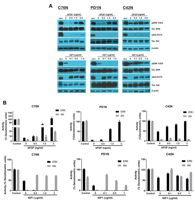
Figure 2. Effect of bFGF and IGF1 on downstream signaling mediators in NCM cells. (A) Cells from C76N, PD1N and C42N were treated or not with increasing concentrations of bFGF (upper panel) and IGF1 (lower panel). Cell lysates were prepared in the presence of protease and phosphatase inhibitors, and equivalent protein was loaded onto each well. Western blots were probed with phosphor-specific antibodies targeted to phospho-ERK1/2, phospho-Akt, total ERK1/2, total Akt and cyclin D. β-actin was used as loading control. (B) Densitometric analysis of relative band intensities using Image J and plotted as percent densitometric units assuming control (HMGS) as one hundred percent.

Figure 2