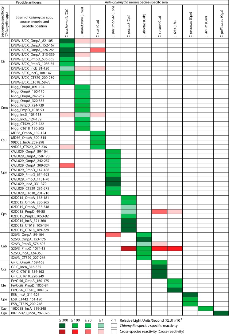
FIG 1 .
Reactivities of 60 peptide antigens from 11 Chlamydia species with Chlamydia species-specific mouse sera. Each peptide was ELISA tested with 11 pools of 9 to 50 hyperimmune mouse sera obtained by 3× intranasal inoculation with live inocula of a single chlamydial species (39). Green cells represent the reactivity of peptide antigens with their corresponding homologous antiserum pools. Red cells indicate peptide antigen cross-reactivity with nonhomologous antisera (ELISA signals > background + 2 standard deviations [SD]). Green and red color intensities indicate signal strength, and white cells indicate nonreactivity. Peptide designations consist of three-letter Chlamydia species acronyms (defined in the headings of columns 3 to 13) followed by strain, source protein, and the amino acid positions of the peptide in the protein. RLU indicates relative light units per second.

