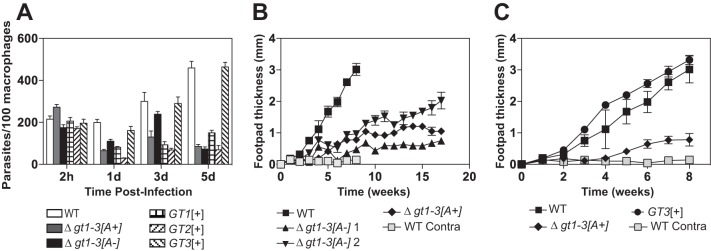
FIG 4 .
Infectivity and virulence of glucose transporter null mutants. (A) Survival of glucose transporter null mutants in primary bone marrow macrophages. Murine macrophages were infected with stationary-phase promastigotes of the indicated parasite lines, and intracellular parasites were enumerated at 2 h and 1, 3, and 5 days after infection. Each sample was counted as 3 separate infections, and the values graphed represent the averages and standard deviations. (B) Lesion size (change in footpad thickness) in BALB/c mice infected with wild-type L. mexicana and Δgt1-3 null mutants that either have ([A+]) or do not have ([A-]) the 29-40k amplicon. Two clonal isolates of the Δgt1-3[A-] line were employed and designated [A-]1 and [A-]2. Five mice each were infected in one hind footpad with 5 × 10⁶ stationary-phase promastigotes of each line. Footpad thickness was measured at the time of infection, and the increase in this value was measured each week. Values plotted represent the average and standard deviations for each cohort of 5 mice. Light gray squares represent measurements of the contralateral noninfected footpad thickness for wild-type parasites. For the Δgt1-3[A+] clone, the infection experiments were repeated a second time with the same results. (C) Lesion size in BALB/c mice infected with wild-type, Δgt1-3[A+], and GT3[+] lines. All data represent a separate experiment from that shown in panel B.

