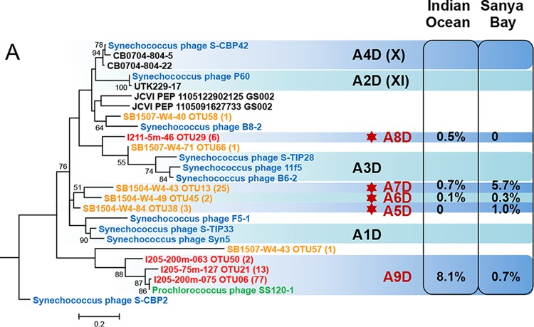
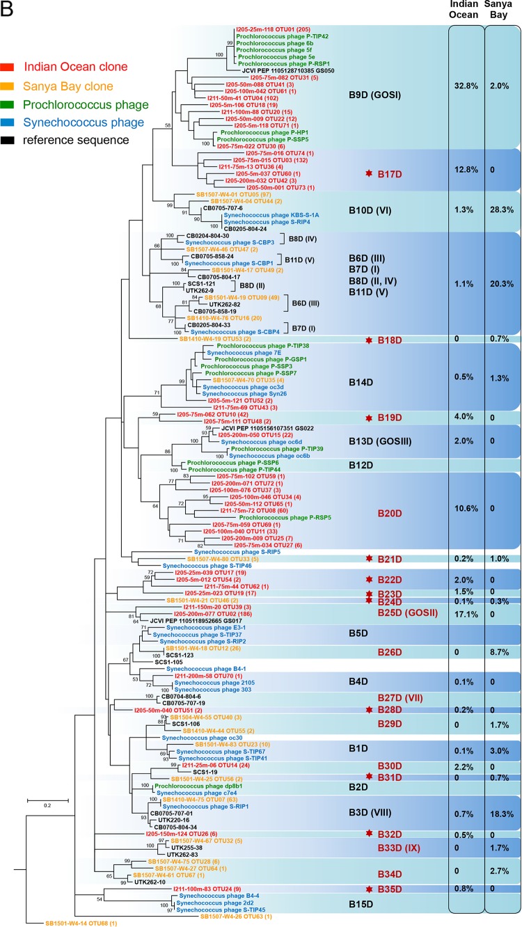
FIG 3.
Maximum likelihood phylogenetic trees based on the viral DNA polymerase protein sequence of cyanopodoviruses. Trees for the MPP-A cluster (A) and the MPP-B cluster (B) were built independently. Environmental sequences obtained in this study are labeled in the format “representative sequence identifier (ID) OTU (no. of sequences assigned to the OTU).” Bootstrap values for 100 sampling replicates are listed only for those higher than 50%. Reference sequence shown in green is from phage isolates infecting Prochlorococcus, those in blue are from phage isolates infecting Synechococcus, and those in black are from environmental clones or from the Global Ocean Sampling metagenomes. Percentages of sequences assigned to each of the genotypes are listed to the right. Genotype labels are according to the numbering by Dekel-Bird and colleagues (30). Genotypes labeled in red indicate those defined in this study, and red star symbols indicate those newly detected here.


