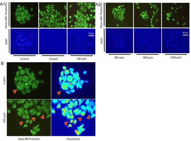
Figure 3.
Immunofluorescent phalloidin/4’,6-diamidino-2-phenylindole (DAPI) staining of HEEpiCs
Note: (A1, 2) The actin cytoskeleton was visualized by staining with Alexa 488 phalloidin (green, the upper panel). Cell nuclei were counterstained with DAPI (blue, the lower panel). The hydrogen-occluding silica microparticles (H2-silica) concentrations were 0 (control), 10, 100, 300, 600, and 1200 ppm. Scale bar: 50 μm (400× magnification). (B) To present the features of Alexa 488 phalloidin more sharply, LUT (look-up table) images in pseudocolor treated with ImageJ were obtained to quantitatively evaluate the number of microvilli.

