Figure 4.
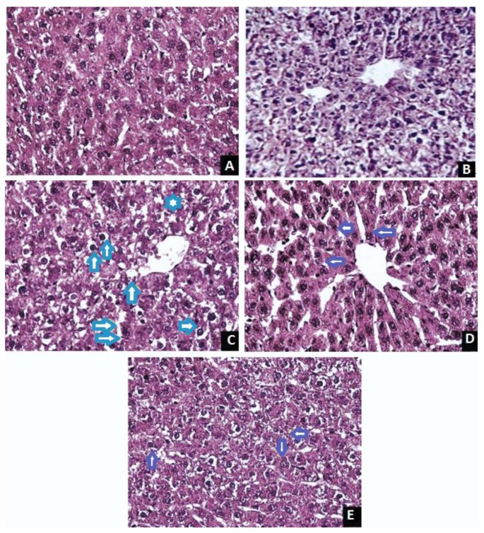
Micrographs showing the effect of Justicia tranquebariensis on TAA-induced hepatic fibrosis in rats. (A) Section of normal control rats showing the histological appearance of hepatocytes with prominent nuclei and cytoplasm (H&E. 400×); (B) Section of J. tranquebariensis (400 mg/kg bw/p.o.) treated control rats showing the histological appearance of hepatocytes with prominent nuclei and cytoplasm (H&E. 400×); (C) Section of TAA (100 mg/kg bw/s.c.) treated control showing fatty degeneration of some hepatocytes, Kupfer cells characterized by cell swelling, the replacement of the cytoplasm with a clear fluid and a centrally located nucleus (↑↑ blue), loss of cell boundaries, hepatic necrosis, collagen and fibronectin deposition and inflammatory cell infiltration (H&E. 400×); (D) Section of TAA (100 mg/kg bw/s.c.) plus J. tranquebariensis (400 mg/kg bw/p.o.) treated rats showing regenerated cells and the almost normal architecture of the liver (↑ violet) with a decrease in collagen and fibronectin deposition (H&E. 400×); (E) Section of TAA (100 mg/kg bw/s.c.) plus silymarin (50 mg/kg bw)-treated rats showing regenerated cells and the almost normal architecture of the liver (↑ violet) with decrease in collagen and fibronectin deposition (H&E. 400×).

