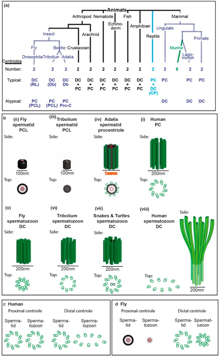
Figure 2.
Spermatozoon typical and atypical centrioles have diverse structures in animals. (a) Phylogenetic tree with representative animal groups. Number of centrioles (first row), which centriole is typical and atypical (second and third rows, respectively), and type of centrioles as well as their modifications (in parenthesis) are indicated; RL, reduced diameter of the centriole lumen; Db, nine doublets of microtubules instead of triplets; PCL, proximal centriole-like structure as depicted in panel “b” or “d”; Pro-c, procentriole; CP, centriole with central pair. (b) Models of spermatid and spermatozoa proximal and distal centriole. The centrioles are depicted from side and top views and are to scale. (c,d) The structural differences between spermatid centrioles and spermatozoa centrioles in human (c) and fly (d).

