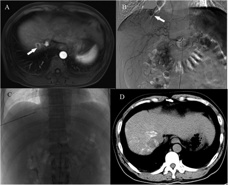
Figure 1.
A, A preprocedure abdominal contrast-enhanced MRI (venous phase) shows a subdiaphragmatic tumor (white arrow). B, Tumor staining is clearly demonstrated by angiography (white arrow). C, An electrode and a microcatheter are in position during the RFA-TACE procedure. D, A noncontrast-enhanced CT scan 3 days after the procedure. CT indicates computed tomography; MRI, magnetic resonance imaging; RFA, radiofrequency ablation; TACE, transarterial chemoembolization.

