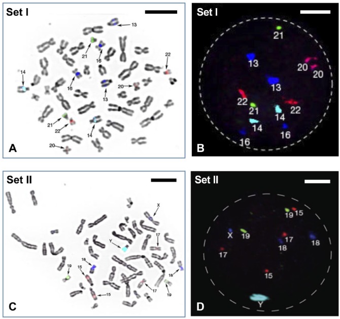
Figure 1.
(A) FISH probe set I (see Table 2) hybridized on one metaphase spread from normal male lymphocyte and (B) one interphase nucleus. Computer-assigned pseudo-colors can be seen showing two copies each of chromosomes 13, 14, 16, 20, 21, and 22. (C) FISH probe set II (see Table 2) hybridized on one metaphase spread from normal male lymphocyte and (D) one interphase nucleus. It showed two copies each of chromosomes 15, 17, 18, 19, and one copy of chromosome X and Y. Scale bars A and C = 5 µm; B and D = 2.5 µm. Abbreviation: FISH, fluorescence in situ hybridization.

