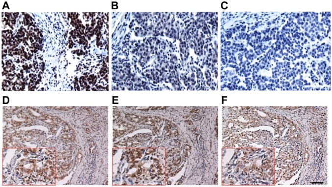
Figure 5.
Antibody specificity determination by peptide competition: (A) Serial sections of CRC-FFPE slides were immunostained with anti-topoI-pS10; (B and C) Slides from the tissue were immunostained with anti-topoI-pS10 in the presence of a phosphopeptide against which the antibody was raised. Two different concentrations: 250 and 500 nm phosphopeptide (Fig. B and C, respectively) were used for this absorption assay. (D) Serial section of gastric cancer FFPE slides were immunostained with anti-topoI-pS10. (E and F) Tissues were also immunostained with anti-topoI-pS10 in the presence of topoI-non-phosphopeptide peptide. Two different concentrations: 250 and 500 nm (Fig. E and F, respectively) were used for the IHC absorption assay. Scale bar = 100 µm. Abbreviations: topoI-pS10, topoisomerase I phosphorylates at serine 10; CRC, colon cancer; FFPE, formalin-fixed paraffin embedded.

