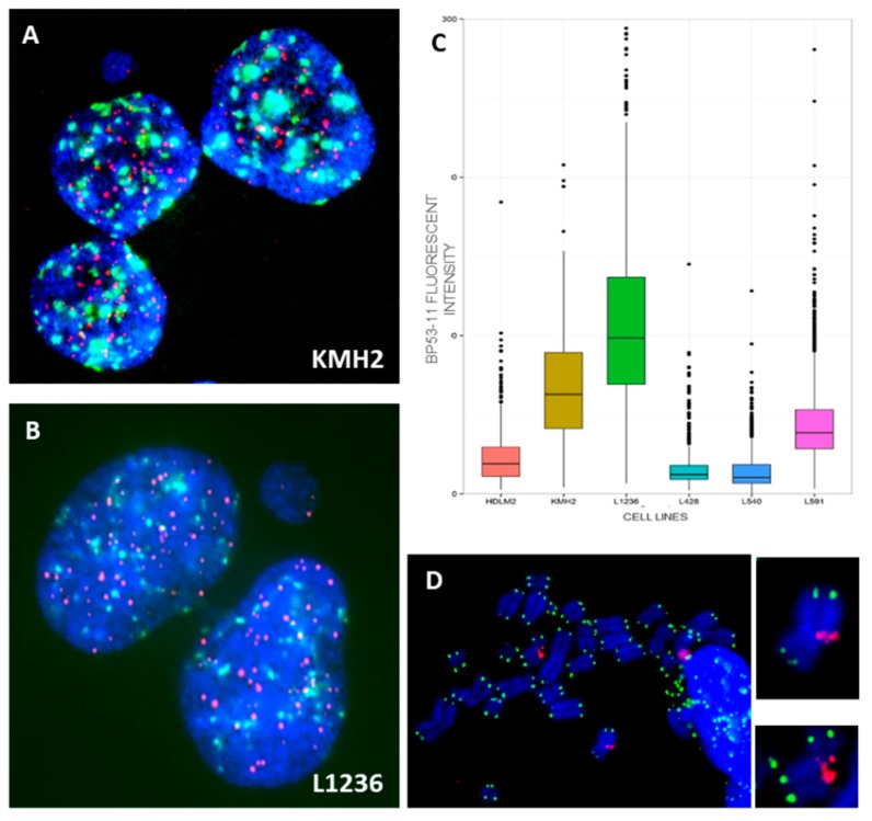
Figure 7.
(A) Small nucleoplasmic bridge without staining connected the daughter cells after four days of culture in the presence of cytochalasin B in KMH2 and (B) in L1236 cells. (C) Total intensity of spontaneous 53BP1 foci in HL cell lines. (D) Representative images obtained following IF-FISH in KMH2 cells showing the presence of γH2AX (red) foci in intra-chromosomal region. Telomeres were stained with green signals (63× magnification).

