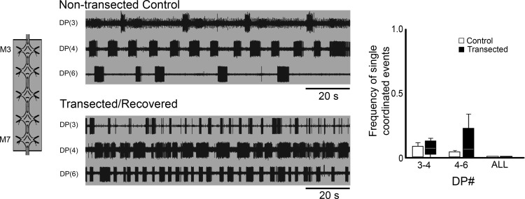
Figure 5.
Dopamine-induced rhythmic activity in M3–M7 ganglion chain preparations of transected and recovered leeches. Left, schematic representation of the preparation showing all ganglia were dopamine-treated (50 µm; gray shading). Traces, extracellular recordings of DP nerve of M3, M4, and M6. Top traces show DP activity from a untransected control, and bottom traces are from an M2/M3 transected and recovered leech. Gray shading denotes dopamine-treated ganglia. Right, boxplots showing the frequency of single DE-3 bursts exhibiting crawl-specific coordination for pairs of DP nerve recordings. White boxes are from nontransected controls and black boxes are from transected/recovered preparations. Lines within the boxes denote the median, and the box range denotes the 25th–75th percentiles. Error bars denote the entire range of the dataset.

