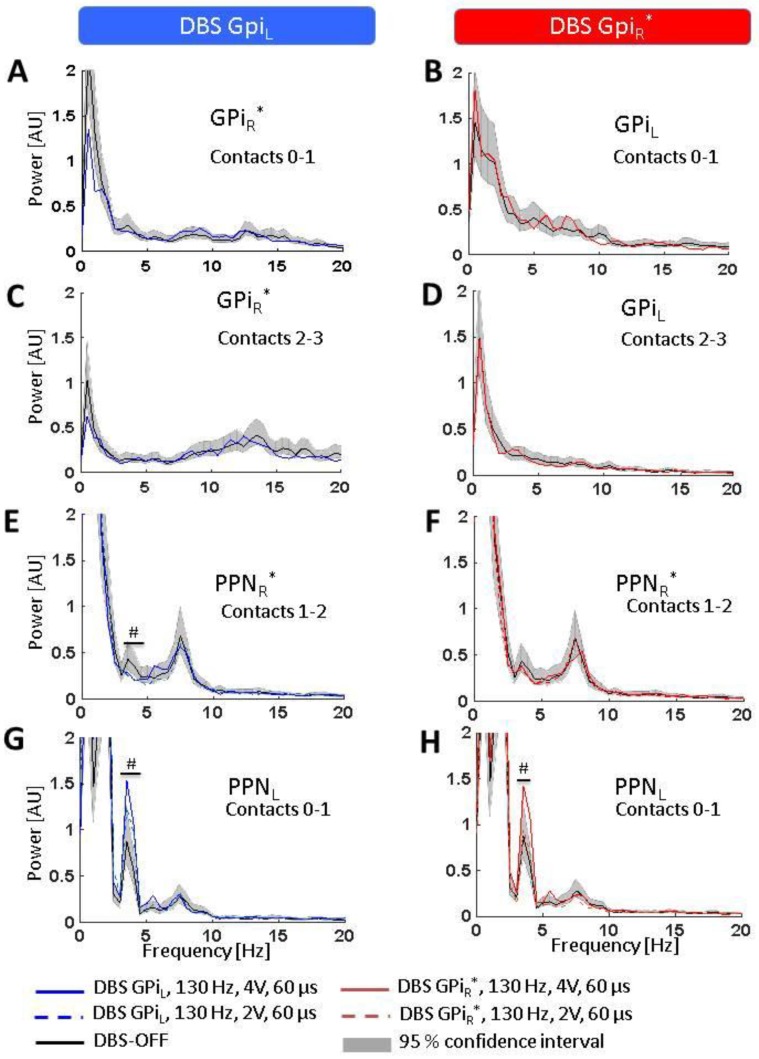
Figure 3.
Effects of GPi-DBS on LFP spectra. Statistical significance levels of differences were derived by comparing the DBS on-spectra to the chi-square distribution assumed for the base line (DBS-OFF) spectra (see Welch, 1967). Significant differences are marked by a ‘#’ indicating a significance level of p < 0.05. (A–D) No spectral changes were observed in GPiR* or GPiL (neither at contacts 0–1 nor 2–3) during DBS of the contralateral GPi; (E,F) A slight but significant decrease was observed in the lower theta band in PPNR* during GPIL-DBS but no spectral change was observed in PPNR* during GPiR*-DBS; (G,H) DBS of either GPiL (left) or GPiR* (right) led to increased activity in the lower theta band in PPNL. Abbreviations: DBS: deep brain stimulation, GPi: globus pallidus internus (L = left, R = right, * denotes clinically more affected side), LFP: local field potentials, PPN: pedunculopontine nucleus region (L = left, R = right, * denotes clinically more affected side).

