Figure 1.
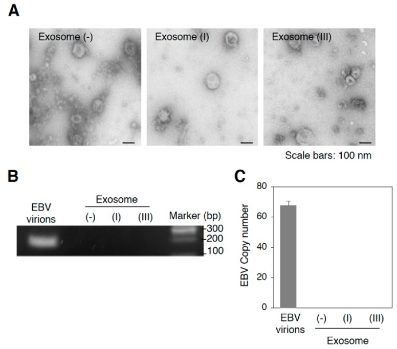
Isolation of exosomes released from Mutu cells. (A) Representative electron micrographs of isolated exosomes from Mutu cells. Isolated Exosome (−), exosome (I), or exosome (III) by ultracentrifugation were analyzed by electron microscopy. Scale bars: 100 nm; (B) Detection of EBV DNA in the isolated exosomes by PCR. DNA was isolated from DNase-treated culture medium containing EBV virions and isolated exosomes followed by PCR. EBV-encoded BALF5 gene was amplified. PCR products were subjected to agarose gel electrophoresis; (C) Detection of EBV DNA in the isolated exosomes by real-time PCR. DNA was isolated from DNase-treated culture medium containing EBV virions and isolated exosomes followed by real-time PCR. EBV-encoded BALF5 gene was amplified. The experiment was performed three times independently and the average and its SD are shown in each condition.

