
Figure 4.
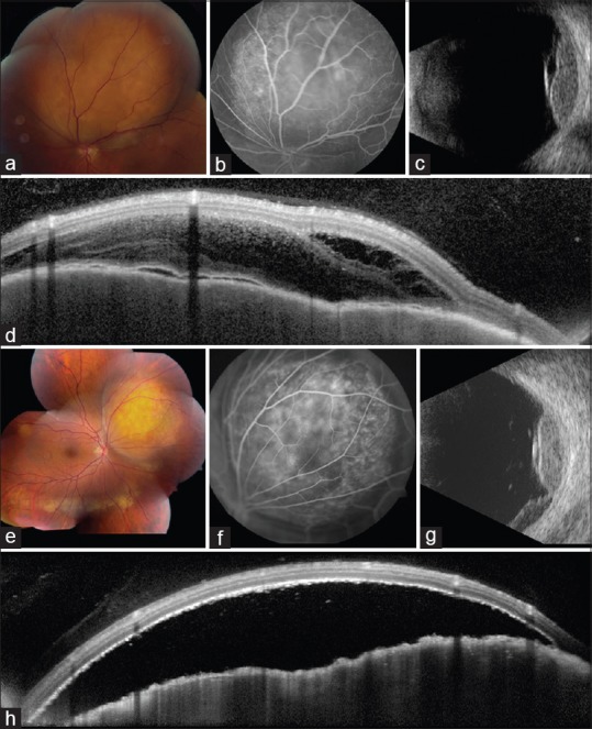
Multimodal imaging in breast and lung cancer metastases. (a-d) Yellow choroidal metastasis (a) from breast cancer, abutting the superior aspect of the optic disc, (b) showing late hyperfluorescence on fluorescein angiography, (c) echodensity with overlying subretinal fluid on ultrasonography, and (d) "lumpy-bumpy" surface with overlying fluid on optical coherence tomography. Yellow choroidal metastasis (e) from lung cancer, (f) showing mottled hyperfluorescence on angiography, (g) echodensity with dependent subretinal fluid on ultrasonography, and (h) "lumpy-bumpy" surface with overlying fluid on OCT