Figure 2.

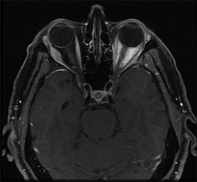
A 51-year-old with a history of metastatic carcinoid tumor with new proptosis. T1 weighted, fat suppressed magnetic resonance imaging shows a right lateral rectus, well-circumscribed mass

A 51-year-old with a history of metastatic carcinoid tumor with new proptosis. T1 weighted, fat suppressed magnetic resonance imaging shows a right lateral rectus, well-circumscribed mass