
Figure 1.
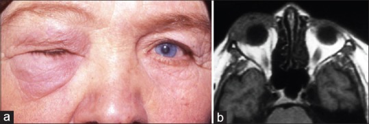
Anterior idiopathic orbital inflammation. (a) A 60-year-old woman presenting with right complete ptosis due to eyelid edema. (b) T1-weighted axial magnetic resonance image shows that inflammation occupying the right anterior orbit has a molded appearance with ill-defined margins and is isointense with respect to extraocular muscles and cerebral gray matter (Reproduced from Gündüz K, Yesiltas YS, Shields CL. Orbital Tumors: A systematic review part II. Expert Rev Ophthalmol 2015;22:485-508)