
Fig. 4.
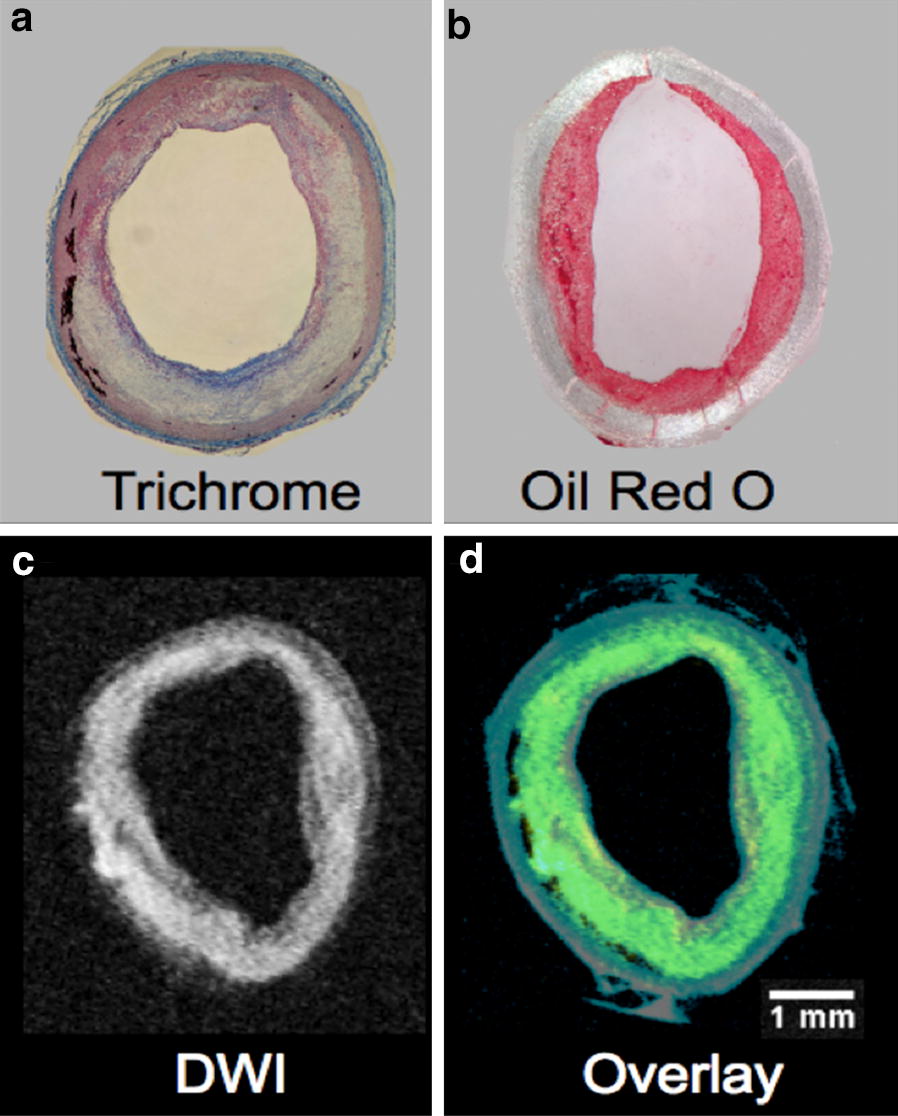
Displays of heterogeneous plaques with abundant lipids in 1% CHOL + injury rabbits by histology and ex vivo MRI. a Masson’s trichrome staining revealed massive heterogeneity with inflammation. b Abundant lipids in the luminal side of the vessel wall were visualized by Oil Red O staining. c Diffusion-weighted MRI (DW-MRI) confirmed substantial cholesteryl ester deposition in the luminal side of the vessel wall of 1% CHOL + injury rabbits [35]. d The DW-MRI sequence was overlaid on the standard T1W image showing all of the components of the vessel wall from 1% CHOL + injury rabbit. DW-MRI is depicted in yellow and T1W in blue. Overlapping regions result in the color green. Scale bar size inset is 1 mm across the images with equivalent scaling (a–d)