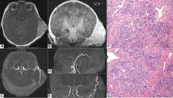
Figure 1.
Preoperative MRI/MRA imaging and tissue histology of a patient with a large congenital scalp hemangioma. (A,B) MR imaging of the brain with and without gadolinium, demonstrating a large extracranial contrast enhancing lesion in the left temporal area; axial and coronal T1-weighted post-gadolinium MR images shown; (C–E) axial, coronal, and sagittal reconstructions of the 3T time-of-flight MR angiogram images, demonstrating the arterial supply of patient's congenital scalp hemangioma arising from superior temporal and occipital arteries; (F,G) 20× images of H&E stains of congenital hemangioma showing dense fibrous stroma with large irregular vessels.

