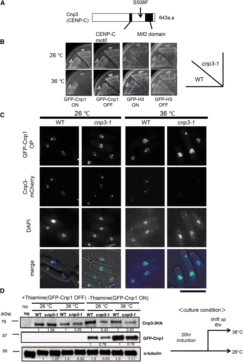
Figure 1.
Distribution of GFP-Cnp1. (A) Schematic illustration of the structure of Cnp3 protein and the position of the cnp3-1 mutation. (B) The wild type strain (WT) and the cnp3-1 mutant (cnp3-1) were transformed with pREP41-GFP- Cnp1 or GFP- H3. The resulting transformants (MS01, MS02, MS03 and MS04) were streaked as indicated and grown at 26°C or 36°C on EMM 2 media with thiamine for repression (GFP-Cnp1 OFF) or without thiamine for derepression (GFP-Cnp1 ON). (C) Localization of GFP-Cnp1 and Cnp3-mCherry were examined in the wild type (MS213) and cnp3-1 cells (MS217). They were grown at 26°C for 20 hr for induction of GFP-Cnp1 in absence of thiamine, and shifted to 36°C for 6 hr in absence of thiamine. The bar indicates 5 μm. (D) The levels of GFP-Cnp1and Cnp3-HA were examined by western blot in the wild type strain (WT: MS525) and the cnp3-1 mutant (cnp3-1: MS527). They were grown as (C) and the samples were taken 20 hr after the induction at 26 °C and 6 hr after the shift to 36 °C. α-tubulin was used as a loading control.

