Figure 4.
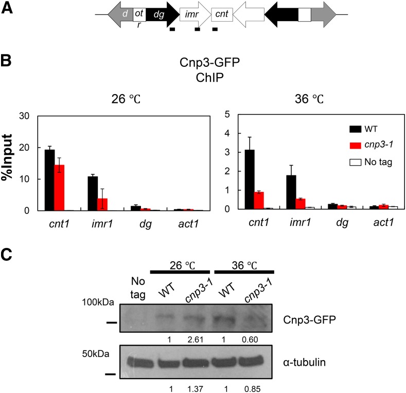
Loss of Cnp3-1 from the centromere. (A) Schematic illustration of the centromere I (Cen I). Each centromere contains a central domain (cnt), flanked by innermost repeats (imr). This core domain is surrounded by arrays of outer repeats (otr). The black bars indicate the position of the primers used in the ChIP analysis. (B) Localization of Cnp3-GFP in Cen I was examined by ChIP analysis in the wild type stain (WT: MS468) and the cnp3-1 mutant (cnp3-1: MS544). The stains were grown at 26 °C and shifted to 36 °C for 6 hr. (C) The level of Cnp3-GFP was examined by western-blot in the wild type strain (WT: MS468) and the cnp3-1 mutant (cnp3-1: MS544) grown as in (B). α-tubulin was used as a loading control.

