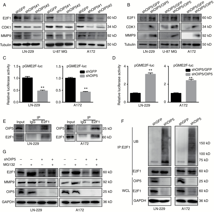
Fig. 5.
OIP5 regulates and stabilizes E2F1 signaling. (A) Protein expression levels of E2F1, CDK1, and MMP9 were analyzed by western blot in LN-229, U-87 MG, and A172 cells expressing shGFP, shOIP5#1, or shOIP5#2. (B) E2F1, CDK1, and MMP9 expression levels of the OIP5-rescued OIP5 knockdown cells. (C) Luciferase assay in LN-229 and A172 cells co-transfected with the reporter plasmids combined with the indicated shRNA. (D) Luciferase assay in OIP5-rescued OIP5 knockdown cells. (E) Interaction of endogenous OIP5 with endogenous E2F1. Equal amounts of LN-229 and A172 cell lysates were used for each immunoprecipitation (IP) reaction. (F) LN-229 and A172 cells expressing shGFP or shOIP5 were treated with MG132 for 6 h before harvesting. Whole cell lysates (WCL) were incubated with anti-E2F1 antibody, and the immunocomplexes were immunoblotted with antibodies against ubiquitin (UB) and E2F1. (G) LN-229 and A172 cells expressing shGFP or shOIP5 were treated with or without MG132 for 6 h before harvesting. Equal amounts of cell lysates were immunoblotted with the indicated antibodies. Data were analyzed using 2-tailed Student’s t-tests, **P < 0.01.

