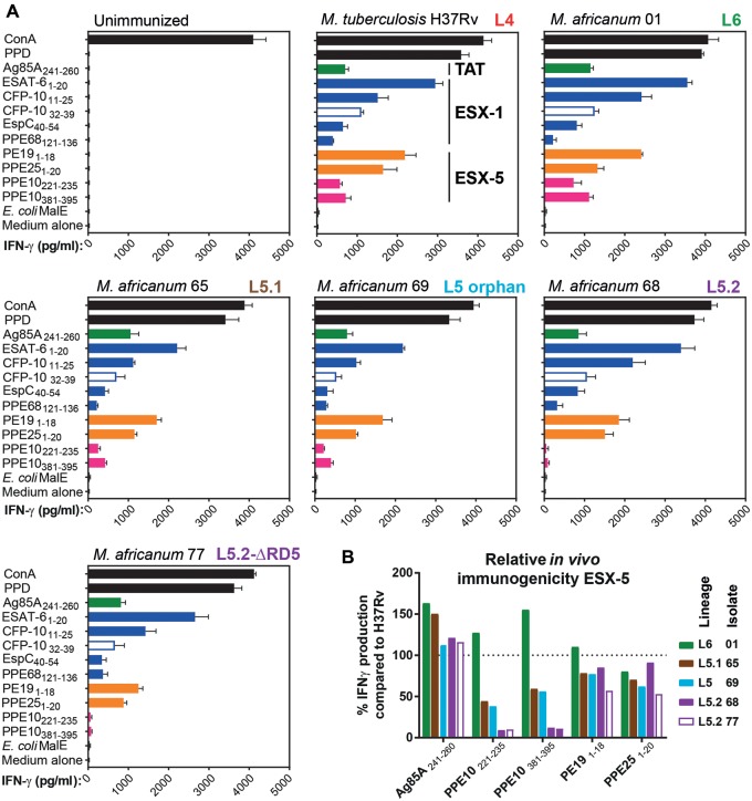
Fig. 4.
—Sublineage-specific differences in immunogenicity of M. africanum. (A) Two 6-week-old female C57BL/6 x CBA F1 (H-2b/k) mice per group subcutaneously received 1×10⁶ CFU/mouse of the indicated mycobacterial strains. Three weeks later, mouse splenocytes were stimulated with peptides containing known immunogenic MHC-II (closed bars) or MHC-I (open bars) restricted epitopes (listed on the y axes). IFN-γ production of stimulated splenocytes was measured as a readout of T-cell stimulation (x axes). The (sub)lineage of the tested strains is indicated in bold typeset in the top-right of each panel. Colors indicate the nature of the tested immunogenic peptides. Green: twin-arginine transported substrate; blue: ESX-1 substrate; orange/pink ESX-5 substrate; pink putative PPE38-dependent substrate; black: positive and negative controls. (B) Relative immunogenicity of ESX-5-secreted substrate PPE10 in M. africanum compared with M. tuberculosis H37Rv (dotted line at 100%), reveals PPE-MPTR secretion defect in L5.2 isolates 68 and 77 (purple bars), while other ESX-5 dependent substrates PE19 and PPE25, or the TAT-secreted substrate Ag85A were not significantly affected.

