Figure 3a:

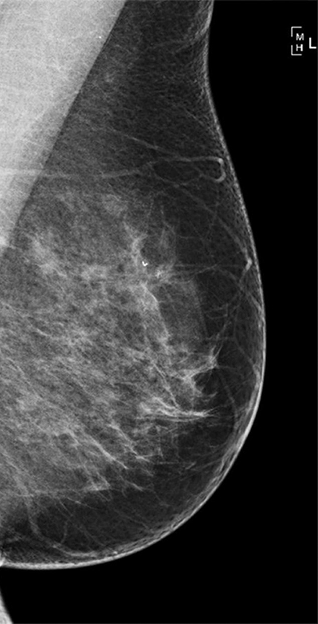
(a–c) Images show a left-sided 6-mm low-grade ductal carcinoma in situ. (a) Left mediolateral oblique mammographic view. (b) Left craniocaudal mammographic view. (c) Left coned magnification of the microcalcification.

(a–c) Images show a left-sided 6-mm low-grade ductal carcinoma in situ. (a) Left mediolateral oblique mammographic view. (b) Left craniocaudal mammographic view. (c) Left coned magnification of the microcalcification.