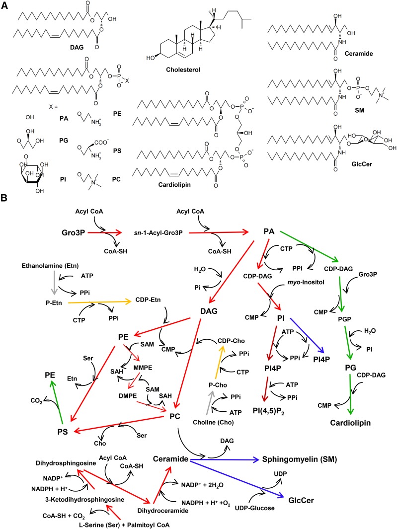
Fig. 1.
Biosynthesis of glycerophospholipids and sphingolipids in mammalian cells. A: The structures of major lipid types in mammalian cells are shown. For simplicity, the diradyl moieties of glycerolipids and sphingolipids are depicted as sn-1-palmitoyl-2-oleoyl (C16:0/C18:1) and N-palmitoyl d18:1-sphingosine (C16-ceramide), respectively, as their typical structures, although variations in diradyl structures occur in cells. B: The biosynthetic pathway of major glycerophospholipids and sphingolipids in typical mammalian cells is shown. Arrow colors represent the organelles in which the reactions predominantly occur: red, ER; orange, nuclear envelope; blue, Golgi apparatus; green, mitochondria; brown, PM; gray, cytosol. Conversion of DAG to PC may also occur in the Golgi apparatus (see text and Fig. 5A). The methylation of PE to produce PC is specific to hepatocytes. Whereas de novo synthesis of PI4P occurs in the Golgi apparatus and PM by different PI-4 kinase isoforms, PI4P of the PM is often converted to PI(4,5)P2 by a PM-localizing PI4P-5 kinase. Gro3P, glycerol-3-phosphate; Pi, inorganic phosphate; PPi, pyrophosphate; P-Cho, phosphocholine; P-Etn, phosphoethanolamine; PGP, PG-1-phosphate; MMPE, N-monomethyl PE; DMPE, N-dimethyl PE; SAM, S-adenosyl- l-methionine; SAH, S-adenosyl- l-homocysteine.

