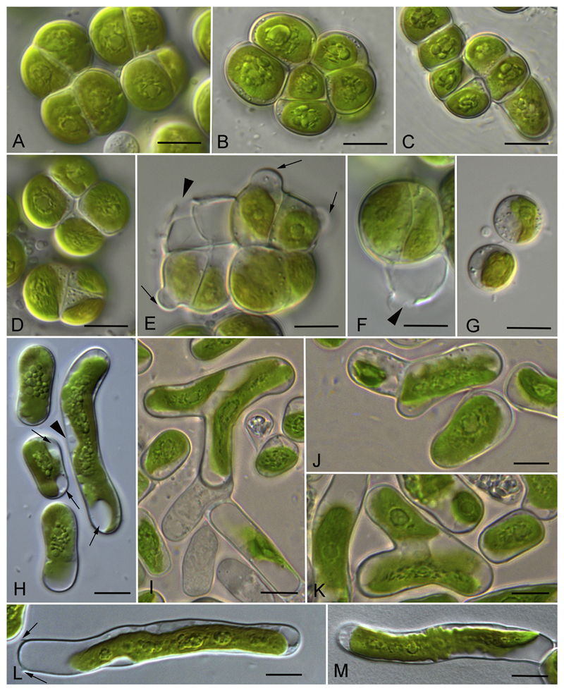
Fig. 5.
Morphology and reproduction of Streptosarcina gen. nov. A–G) S. arenaria sp. nov. (AL-63 (B, C) and Prim-3-3 (A, D-G). A, B, D). Packet-like vegetative thallus. C) Filaments. E, F) Formation of sporangia (arrows) and empty sporangia with openings in cell wall (arrowheads). G) Stopped zoospores. H–M) S. costaricana sp. nov. (SAG 36.98). H) Unicellular stage, nucleus (arrowhead) and terminal vacuoles (arrows) are visible. I-K) Branching of filaments. L, M) Elongated cells with multiple pyrenoids from old culture, H-like fragments of cell wall are visible (arrows). Scale bars are 10 μm.

