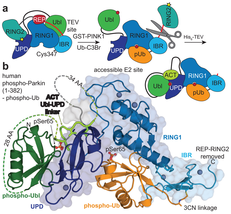
Figure 2. Structure of the phosphorylated Parkin core.
a, Schematic for obtaining a crystallisable phosphorylated Parkin core. Scissors indicate the introduction of a tobacco etch virus (TEV) protease cleavage site after the IBR domain (aa 382). b, Crystal structure at 1.80 Å of the human phosphorylated Parkin core lacking RING2, bound to phospho-ubiquitin. Phosphorylated residues are shown in ball-and-stick representation. A cartoon representation akin Fig. 2a is shown to the right. Also see Extended Data Fig. 6 and Extended Data Table 1.

