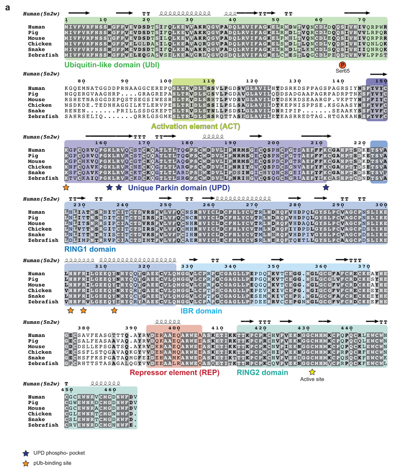
Extended Data Figure 4. A conserved linker between Ubl and UPD.
Sequence alignment of Parkin, with domains coloured corresponding to 5n2w 6 as in Extended Data Fig. 1. Phosphate binding pockets are labelled. The linker region between Ubl and UPD (aa 76-143) contains two strings of highly conserved residues. Residues upstream and downstream of the conserved region are unconserved both in sequence and linker length. Thamnophis sirtalis (Ts, garter snake) Parkin shows the smallest number of residues in the linker (upstream, 25 aa in human Parkin, 18 aa in TsParkin; downstream, 18 aa in human Parkin, 11 aa in TsParkin). See also Extended Data Fig. 8d.

