. 2018 May 18;126(5):057005. doi: 10.1289/EHP2387

Figure 6.

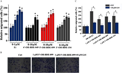
Figure 6A is a bar graph plotting the percentage of relative migrated cells (y-axis) across concentrations of E2 in 0 to 1 micromolar, and 4 prime OH BDE 049, 5 prime OH BDE 099, and 3 prime OH BDE 154 (x-axis), all measured in 0 to 10 micromolar. Figure 6B presents micrographs of the Boyden chamber assay for the control group, 1 micromolar 5 prime OH BDE 099 and 1 micromolar 5 prime OH BDE 099 plus 10 micromolar G15. Figure 6C is a bar graph plotting the percentage of relative migrated cells (y-axis) across control group, 100 nanomolar E2, 1 micromolar 4 prime OH BDE 049, 1 micromolar 5 prime OH BDE 099, and 1 micromolar 3 prime OH BDE 154 (x-axis) in the presence of 10 micromolar G15 and in the absence of G15.

Effects of 17β-estradiol (E2) and three hydroxylated polybrominated diphenyl ethers (OH-PBDEs) on SKBR3 cell migration detected by Boyden chamber assay and the inhibitory effects of G15. (A) SKBR3 cell migration induced by different concentrations of E2 (0, 0.1nM, 1nM, 10nM, 100nM, and 1μM) and OH-PBDEs (0, 1nM, 10nM, 100nM, 1μM, and 10μM). (B) A typical Boyden chamber assay result for 1μM 5ʹ-OH-BDE-099 in the absence or presence of 10μM G15, a G protein–coupled estrogen receptor antagonist. (C) SKBR3 cell migration induced by 100nM E2 and 1μM OH-PBDEs in the absence or presence of 10μM G15. The relative migration of cells is calculated by setting the count of migrated cells of the control group (Ctrl) as 100%. *p<0.05 compared with the control group (Ctrl, 0.1% dimethyl sulfoxide). #p<0.05 compared with the groups treated with compounds in absence of G15.