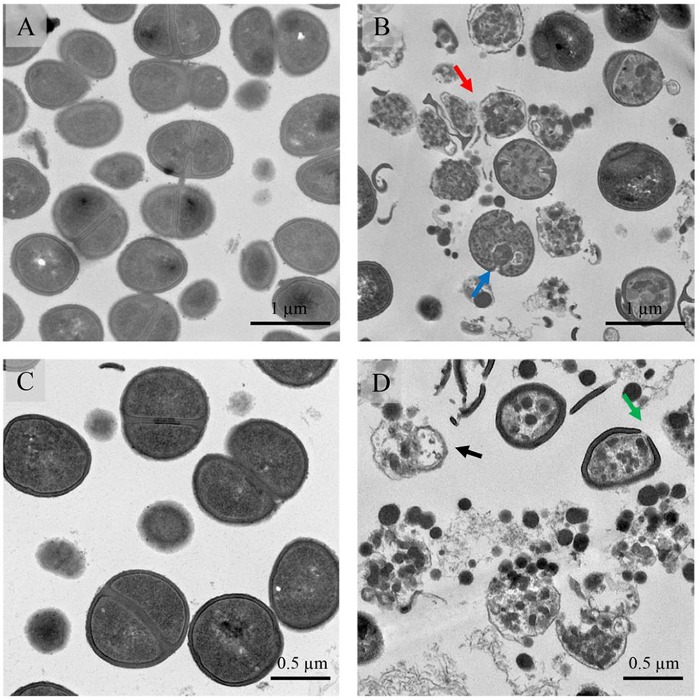
Fig 5. TEM micrographs of S. epidermidis after treatment with BcDef1.
(A and C) Untreated cells possessed intact cell walls and cell membranes. After 2 h of BcDef1 treatment, (B and D), BcDef1 induced cell lysis (black arrow) and ultrastructural damage in S. epidermidis cells, including some mesosome-like structure formation (blue arrow), and ruptures of cell walls (green arrow) and cell membranes (red arrow).

