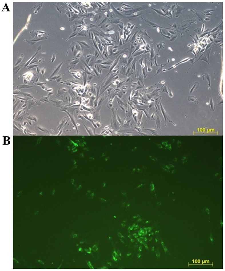
Figure 1.

Representative fluorescent images of co-cultured ECs. Endometriotic ECs were unstained and healthy ECs were stained with Calcein AM 24 h after cell seeding. Calcein AM-stained cells are visualized in green. (A) Blank channel. (B) Green channel. Scale bar, 100 µm. ECs, endometrial cells.
