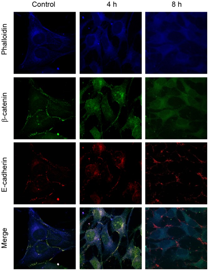
Figure 4.
Immunofluorescence analysis of β-catenin and E-cadherin in Saos-2 osteoblastic cells following tensile stress. β-catenin was localized on the cytomembrane in the control group and in the cytoplasm and nucleus after 4 or 8 h of 12% tensile stress. However, E-cadherin was distributed on the cytomembrane throughout. Magnification, ×600.

