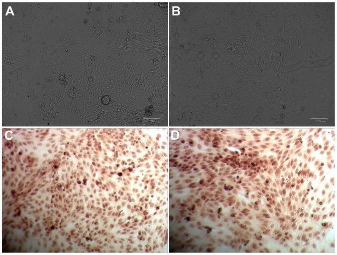
Figure 1.
Morphology characterization of primary renal tubular epithelial cells. Light microscopy images captured following culture for (A) 36 h and (B) 7 days. Immunohistochemical images captured following culture for (C) 36 h and (D) 7 days. At 36 h time intervals, the epithelioid cell population presented with an ‘island-like’ distribution; and at 7 day time intervals, cells were distributed as typical multilateral cobblestones with tight arrangements and high transparency. Brown granules exhibited non-uniform and scattered distributed in the cytoplasm and perinuclear region of cells. Magnification, ×20.

