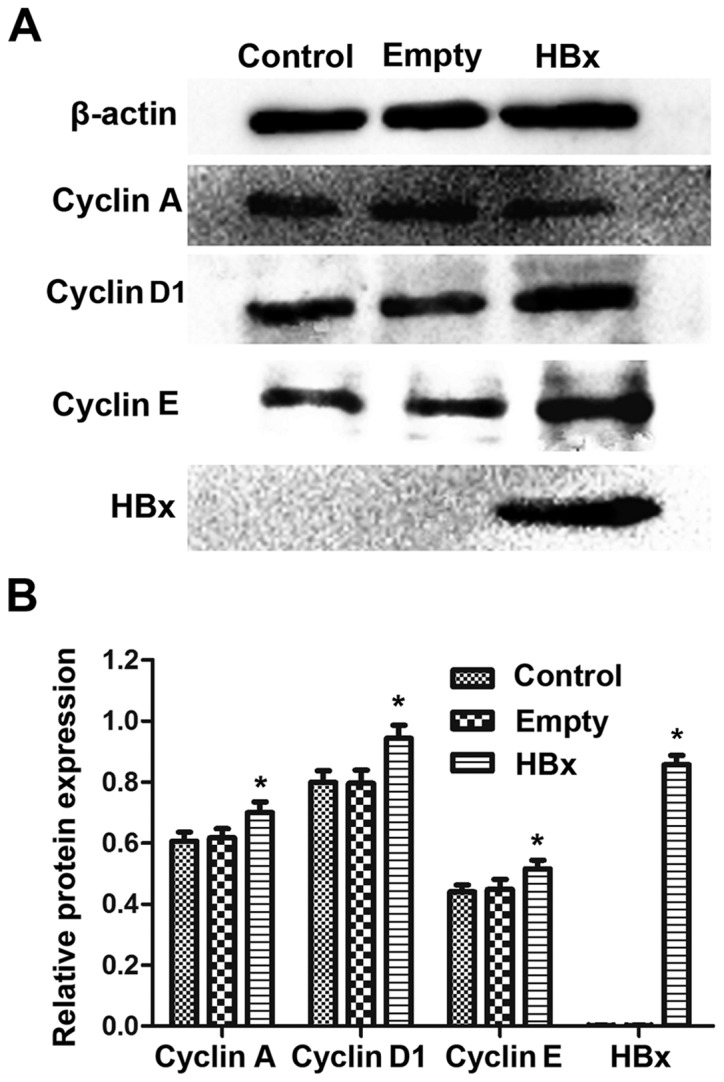
Figure 3.

Protein levels of HBx, cyclin A, cyclin D1 and cyclin E in renal tubular epithelial cells in the control, empty and HBx groups were assessed western blot analysis. (A) Representative western blot bands for cyclin A, cyclin D1, cyclin E and HBx in control, empty and HBx groups are presented. (B) Densitometric analysis was performed to quantify the protein expression levels and perform statistical analysis. Cells transfected with empty plasmid pcDNA3.1(+) and recombinant plasmid pcDNA3.1(+)-HBx were designated as the empty and HBx groups, respectively. Cells without transfection served as the control group. *P<0.05 vs. empty group. HBx, hepatitis B virus X protein.
