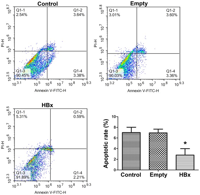
Figure 4.
Cell apoptosis of renal tubular epithelial cells in the control, empty and HBx groups was evaluated by Annexin V-FITC/PI double staining and subsequent flow cytometry. Cells transfected with empty plasmid pcDNA3.1(+) and recombinant plasmid pcDNA3.1(+)-HBx were designated as the empty and HBx groups, respectively. Cells without transfection served as the control group. Q1-2 and Q1-4 quadrants represent cells undergoing apoptosis. *P<0.05 vs. empty group. HBx, hepatitis B virus X protein; FITC, fluorescein isothiocyanate; PI, propidium iodide.

